

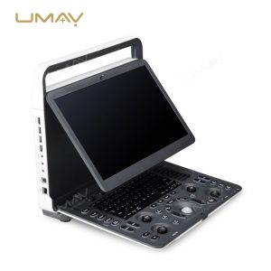
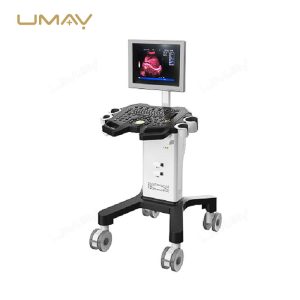


Ultra-thin B/W ultrasound machine with real-time spatial compound imaging
Quick Info.
- SKU NO.: UMY-US-102
- Device Classification: Class Ⅱ
- Warranty: 1 Year
- Power Source: Electric
- Transport Package: Carton or Wooden Box
- Origin: China
- Material: Metal, Plastic
- After-Sale Service: Online Technical Support
- Production Capacity: 3600 Sets/Year
The Ultra-thin B/W Ultrasound Machine offers real-time spatial compound imaging for enhanced diagnostic accuracy. Its compact design ensures easy portability without compromising on performance. Ideal for imaging abdominal, musculoskeletal, and cardiovascular conditions, it provides clear, detailed images to support quick decision-making. The real-time imaging feature allows for precise monitoring of organ conditions, vascular health, and more. This ultrasound machine is a reliable tool for healthcare professionals seeking a space-efficient, high-performance solution for a variety of clinical applications.
Advanced Imaging Technologies:
- REAL-TIME Spatial Compound Imaging
- 3D Free Hand Rebuilding Software (Recommended Option)
- Inversion Harmonic Imaging
- Speckle Reduction & Edge Enhancement
Versatile Scanning Capabilities:
- Multiple Scan Modes B, B+B, 4B, B+M, PW, THI
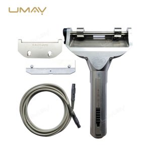
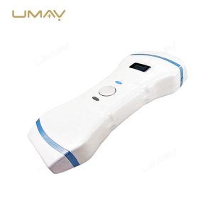
- Support Convex/Linear/Cavity/Micro-convex/Rectal Probes
Automated Measurement Functions:
- Automatic Vascular Intima Measurement
- Automatic Envelope Spectrum Measurement
- Multiple Application and Software Measurement Packag
Core Hardware Specifications:
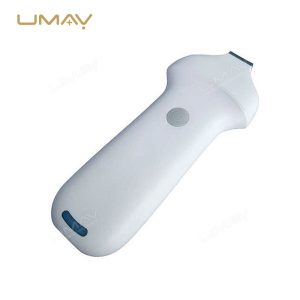
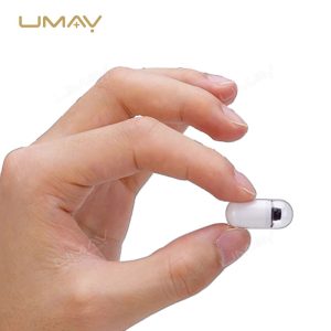
- Light and Thin Design
- 15.6 inches 1920×1080 Resolution HD Display
- Built-in Battery Lasts More Than 4 hours
- 2 Transducer Ports
- Quick Knob of Menu Parameter Adjust
The Specific Parameters
| ITEMS | PARAMETERS |
|---|---|
| Gray-Scale Imaging Mode Performance | |
| Probe Types & Models | Micro-convex (3.5C8020G-2), Convex (3.5C8060G-2), Cavity (6.5C8010G-2), Linear Array (7.5L8040G-2) |
| Nominal Frequency (MHz) | 3.5, 3.5, 6.5, 7.5 |
| Scan Depth (mm) | ≥140, ≥160, ≥40, ≥50 |
| Lateral Resolution (mm) | ≤3 (depth≤80), ≤4 (80<depth≤130); ≤3 (depth≤80), ≤4 (80<depth≤130); ≤2 (depth≤30), ≤2 (depth≤40) |
| Axial Resolution (mm) | ≤2 (depth≤80), ≤2 (depth≤80); ≤3 (80<depth≤130); ≤1 (depth≤40), ≤1 (depth≤50) |
| Blind Area (mm) | ≤7, ≤5, ≤4, ≤3 |
| Transverse Geometry Precision (%) | ≤20, ≤15, ≤10, ≤10 |
| Longitudinal Geometric Location Accuracy (%) | ≤10, ≤10, ≤5, ≤5 |
| Slice Thickness (mm) | ≤5, ≤5, ≤5, ≤5 |
| Perimeter & Area Measured Deviation (%) | ≤±20, ≤±20, ≤±20, ≤±20 |
| M Mode Time Display Error (%) | ≤±10, ≤±10, ≤±10, ≤±10 |
| Color Doppler Imaging Mode Performance | |
| Investigation Depth (Color Blood Flow Mode) | ≥90mm, ≥100mm, ≥40mm, ≥50mm |
| Investigation Depth (Doppler Spectrum Mode) | ≥90mm, ≥100mm, ≥40mm, ≥50mm |
| Blood Flow Speed Reading Error | ≤±15% |
| Doppler Spectrum Mode Performance | |
| Investigation Depth (Color Blood Flow Mode) | ≥90mm, ≥100mm, ≥40mm, ≥50mm |
| Investigation Depth (Doppler Spectrum Mode) | ≥90mm, ≥100mm, ≥40mm, ≥50mm |
| Blood Flow Speed Reading Error | ≤±15% |
| Imaging Mode | |
| B, B+B, 4B Mode | Supported |
| B+M, M Mode | Supported |
| CFM Color Flow Mode | Optional |
| B+CFM Mode | Optional |
| B+CFM+PW Triple Mode | Optional |
| PDI Power Doppler Mode | Optional |
| B+PDI Mode | Optional |
| PW Mode | Supported |
| Power Source Requirement | |
| Input Voltage | AC 100V-240V |
| Frequency | 50Hz/60Hz |
| Output Voltage | 19V ± 1V |
| Power Consumption | 50VA |
| Overall Dimensions | |
| Device | 378mm × 350mm × 56mm |
| Net Weight | Approximately 4Kg |
| Package Size | 445mm × 185mm × 435mm |
| Aluminum Box Package Size | 445mm × 185mm × 435mm |
| Gross Weight | 6.2Kg |
| Aluminum Box Gross Weight | 9Kg |
| Product Lifespan | 6 years (excluding the battery) |
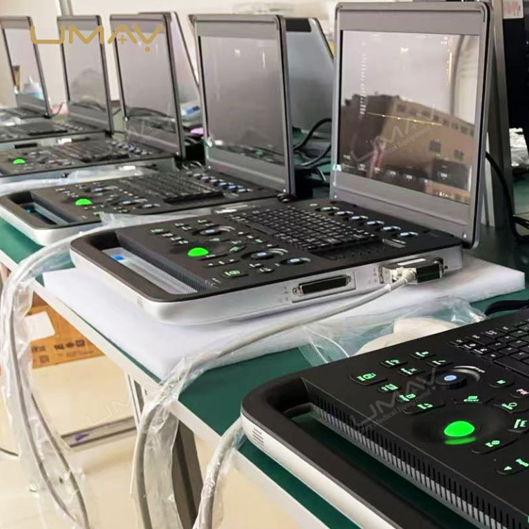
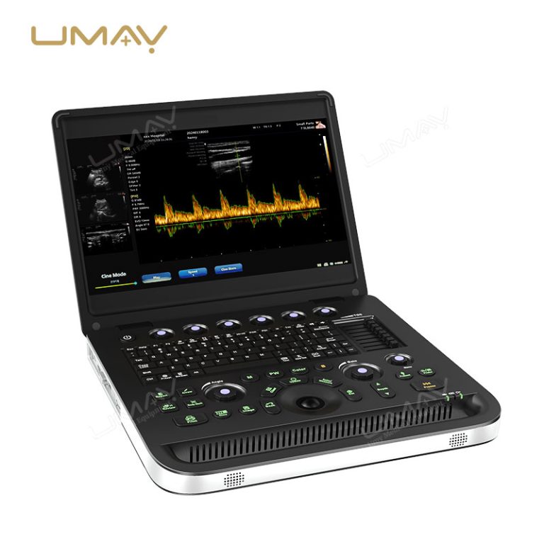
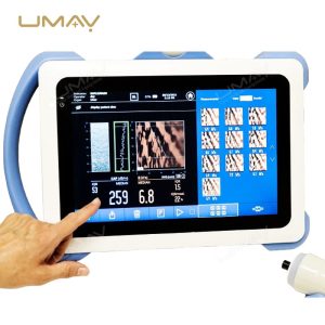
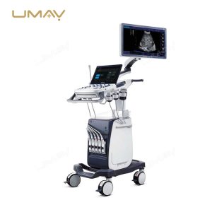
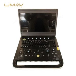
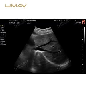
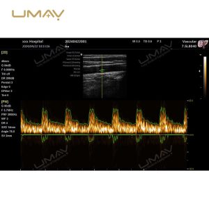
It is showing spectral Doppler imaging with waveform analysis. This model supports multiple imaging modes including B, B+B, 4B, B+M, M modes, with optional CFM Color Flow, PDI Power Doppler, and PW capabilities. The specialized control panel features dedicated knobs and function keys for precise adjustments during examinations. The display shows real-time ultrasonic waveforms with corresponding diagnostic data, demonstrating the system’s clinical capabilities for vascular and cardiac assessments. The ergonomic design integrates advanced imaging technology into a compact, portable workstation.

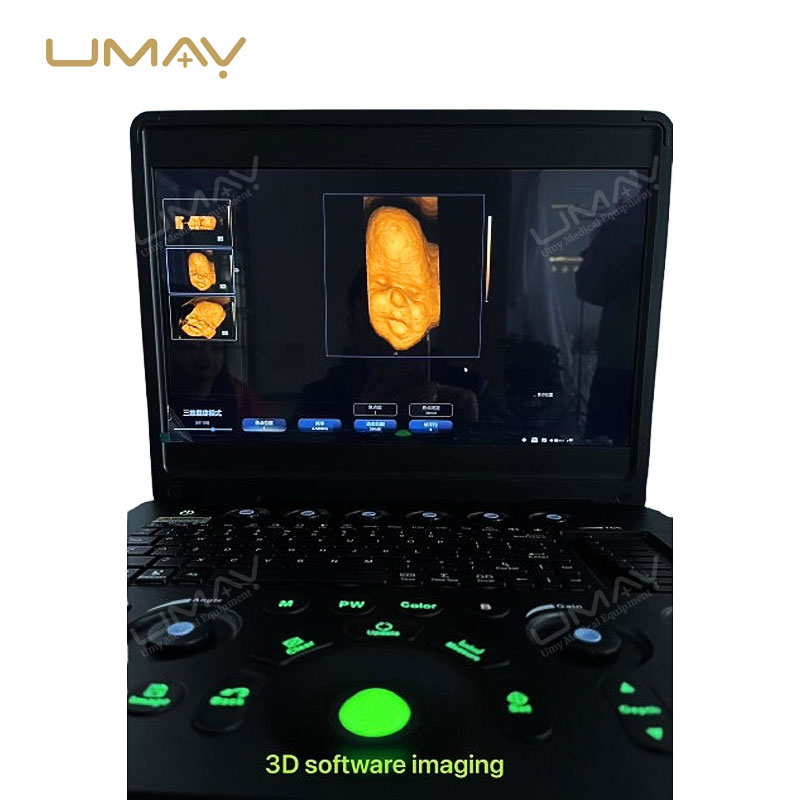
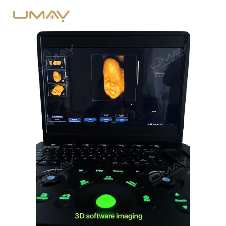
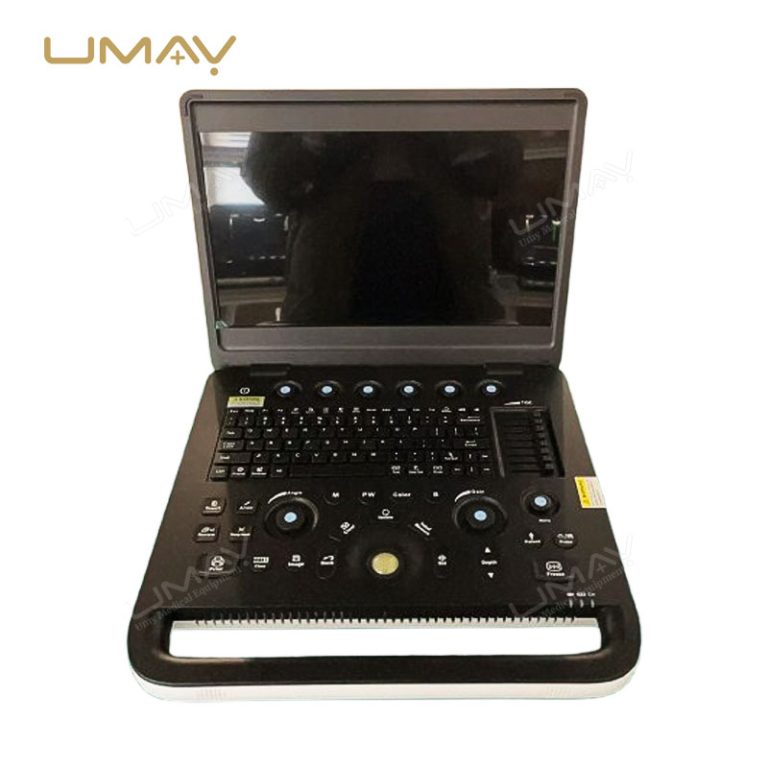
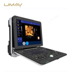
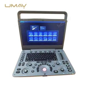
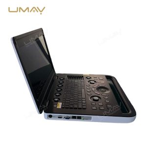
The UMY ultrasound system displaying its 3D imaging software capabilities with a fetal ultrasound image on screen. The device features specialized keyboard controls with illuminated green function keys for intuitive operation in darkened examination rooms. The image demonstrates the system’s advanced visualization technology, capable of rendering detailed 3D representations for comprehensive prenatal examinations. The laptop-style design combines powerful imaging software with user-friendly controls, making complex diagnostic procedures more accessible for healthcare providers.
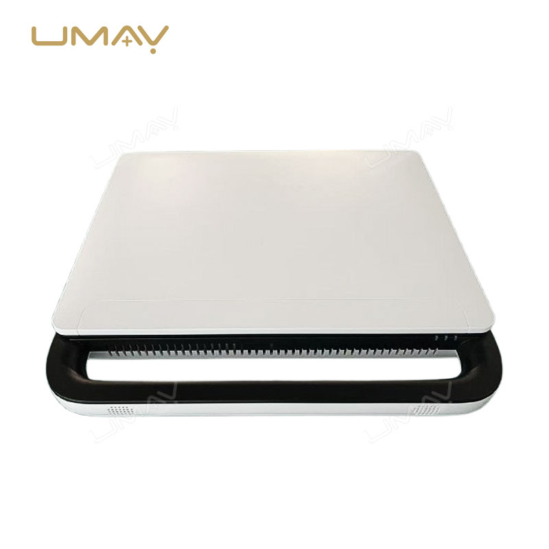
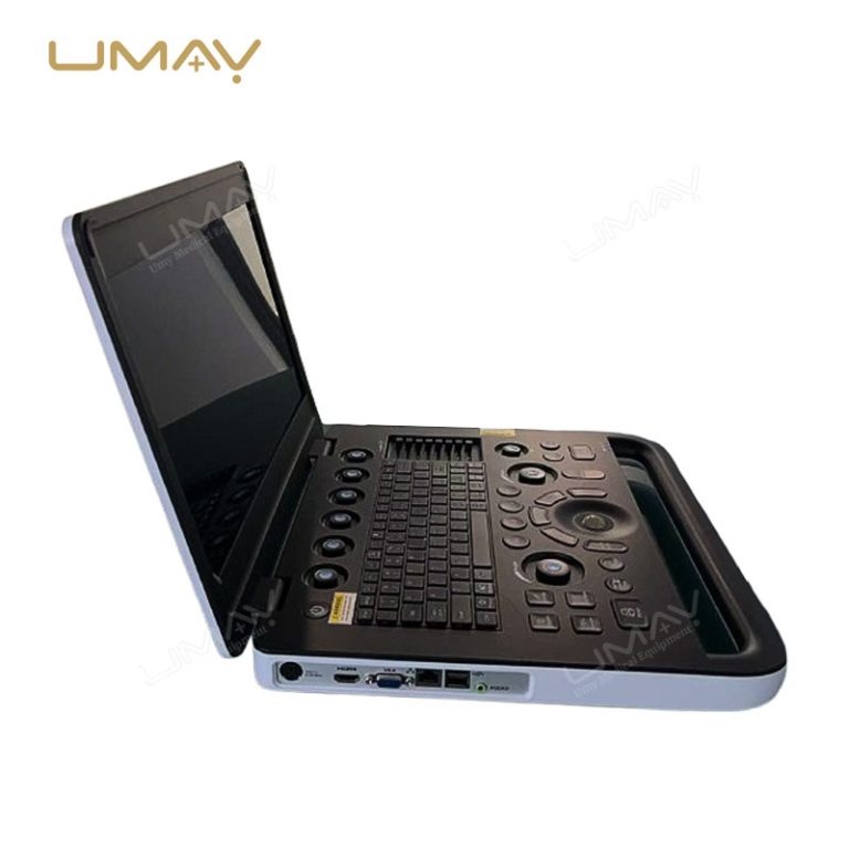
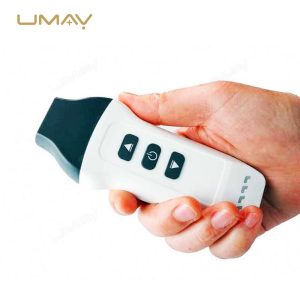
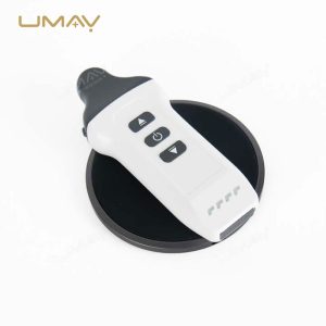
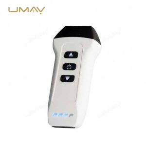
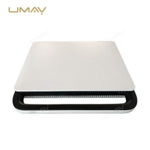
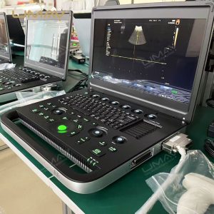
A sleek, compact design with a white top surface and black base. This lightweight diagnostic tool weighs only 4kg and measures 378mm × 350mm × 56mm, making it highly portable for medical professionals. The streamlined profile includes ventilation along the back edge and a handle design for easy transport between examination rooms or medical facilities. Ideal for clinics with space constraints or professionals who need diagnostic capabilities on-the-go.