Radiology plays a vital role in modern medicine, offering advanced imaging techniques that help diagnose and monitor a wide range of medical conditions. Familiarizing yourself with the terminology used in radiology can improve communication between patients and healthcare providers, making the diagnostic process less intimidating and more collaborative.
Today, we’ll take a closer look at some of the most important terms in radiology. These insights will help you better understand radiology procedures, reports, and the critical role imaging plays in healthcare.
-
Computed Tomography (CT Scan)
Did You Know? CT scans can produce images of internal organs in just minutes, making them invaluable for emergency medicine.
Application: They are commonly used to quickly diagnose conditions such as internal bleeding or organ injuries after trauma.
-
Magnetic Resonance Imaging (MRI)
Did You Know? MRI uses strong magnets and radio waves instead of ionizing radiation, making it safer for repeated use, especially in children and pregnant women.
Application: It is frequently used to evaluate soft tissue injuries, brain disorders, and joint problems.
-
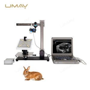
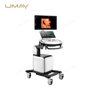
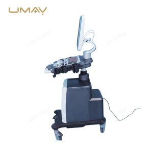
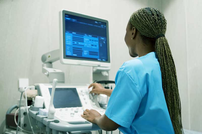
Ultrasound (US)
Did You Know? Ultrasound is safe for monitoring fetal development during pregnancy because it does not use ionizing radiation.
Application: It is commonly used for prenatal check-ups and assessing abdominal organs like the liver and kidneys.
-
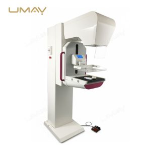
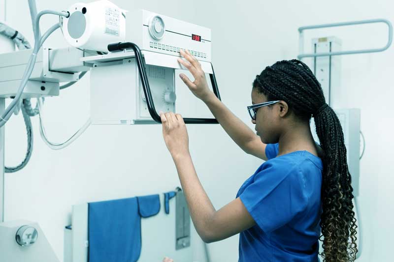
X-ray
Did You Know? X-rays were first discovered in 1895 by Wilhelm Conrad Roentgen, revolutionizing medical diagnostics by allowing visualization of bones and internal structures without surgery.
Application: They are widely used for diagnosing fractures, infections, and dental issues.
-
Angiography
Did You Know? Angiography can help detect blockages or abnormalities in blood vessels, which is crucial for diagnosing conditions like heart disease or aneurysms.
Application: Patients undergoing angiography may have this test to assess blood flow before procedures like stent placements.
-
CT Angiogram
Did You Know? A CT angiogram combines CT imaging with contrast material to visualize blood vessels and assess conditions like aneurysms or blockages.
Application: This non-invasive procedure is often used in emergency settings to quickly evaluate vascular issues.
-
Biopsy
Did You Know? A biopsy can be performed under imaging guidance (like ultrasound or CT) to ensure accurate targeting of abnormal tissues.
Application: This technique is often used to diagnose cancer by obtaining tissue samples from suspicious lesions.
-
Bone Densitometry
Did You Know? Bone densitometry is a key test for assessing osteoporosis risk, especially in postmenopausal women.
Application: Patients may undergo this test as part of routine screenings to prevent fractures.
-
Fluoroscopy
Did You Know? Fluoroscopy allows real-time visualization of moving body parts, such as the digestive tract during swallowing studies.
Application: It is often used in procedures like placing a catheter or guiding injections into joints.
-
Radiolucent
Did You Know? Structures that are radiolucent appear darker on X-ray images because they allow more radiation to pass through them, such as air-filled spaces in the lungs.
Application: Understanding this concept helps radiologists distinguish between different types of tissues and fluids on imaging studies.
-
Radioopaque
Did You Know? Radioopaque materials appear white on X-rays because they block radiation; this property is utilized in contrast agents for clearer imaging results.
Application: Contrast agents are often used during X-rays or CT scans to enhance visibility of blood vessels or organs.
-
Radiopharmaceuticals
Did You Know? Radiopharmaceuticals are radioactive compounds used in nuclear medicine to target specific organs or tissues for imaging or treatment purposes.
Application: Patients undergoing a thyroid scan receive a radiopharmaceutical to evaluate thyroid function and detect abnormalities.
-
Attenuation
Did You Know? Different tissues attenuate X-rays at varying rates, allowing radiologists to differentiate between healthy and diseased tissues.
Application: This principle is fundamental in CT scans, where different densities create detailed images of internal structures.
-
Tomography
Did You Know? Tomographic techniques like CT and MRI provide cross-sectional images that help visualize complex structures within the body more clearly than traditional X-rays.
Application: These techniques are essential for diagnosing cancers, brain disorders, and internal injuries.
-
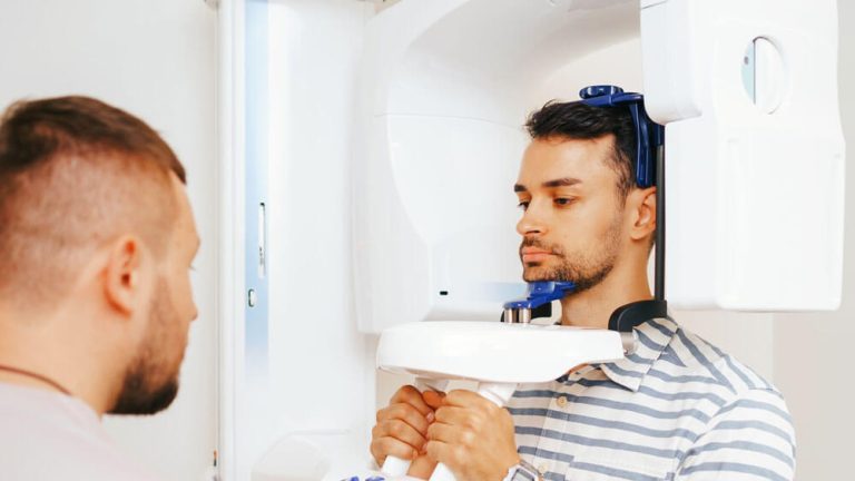
Oblique View
Did You Know? An oblique view in radiology provides a different angle of examination, which can reveal structures that might be obscured in standard views.
Application: This view is frequently used in dental X-rays to assess tooth alignment and detect cavities.
-
Anteroposterior (AP)
Did You Know? The AP view is commonly used in chest X-rays to provide a clear image of the heart and lungs.
Application: This position is essential for diagnosing pneumonia or heart enlargement.
-
Lateral
Did You Know? Lateral views are crucial for assessing the alignment of bones and detecting fractures that may not be visible in frontal views.
Application: This view is commonly used in orthopedic evaluations.
-
Lesion
Did You Know? Not all lesions are cancerous; many can be benign or related to other conditions like infections or inflammation.
Application: Radiologists analyze the characteristics of lesions on imaging studies to guide further testing or treatment.
-
Hematuria
Did You Know? Hematuria, or blood in urine, can be evaluated using imaging studies like ultrasound or CT scans to identify potential causes such as kidney stones or tumors.
Application: Patients presenting with hematuria may undergo these imaging tests as part of their diagnostic workup.
-
Myelogram
Did You Know? A myelogram involves injecting contrast dye into the spinal canal, allowing for better visualization of the spinal cord and nerve roots on X-rays or CT scans.
Application: This test is often used to diagnose conditions like herniated discs or spinal stenosis.
-
Radiographer
Did You Know? Radiographers play a vital role in patient care by ensuring comfort and safety during imaging procedures while obtaining high-quality images for diagnosis.
Application: They interact with patients directly during X-ray and MRI exams, explaining procedures and addressing concerns.
-
Radiologist
Did You Know? Radiologists often collaborate with other specialists to determine the best course of action based on imaging findings, enhancing patient care outcomes.
Application: They interpret images from various modalities to diagnose conditions ranging from fractures to tumors.
-
Nuclear Medicine
Did You Know? Nuclear medicine scans can provide information about organ function as well as structure, which is unique compared to other imaging modalities.
Application: Patients may receive a PET scan to assess cancer spread through metabolic activity.
-
Interventional Radiology
Did You Know? Interventional radiology allows doctors to perform minimally invasive procedures using imaging guidance, reducing recovery time compared to traditional surgery.
Application: Patients might have procedures like catheter placements or biopsies performed by interventional radiologists.
-
Supine & Prone
Did You Know? The terms supine and prone refer to patient positioning during imaging; supine means lying on the back, while prone means lying on the stomach.
Application: Depending on the area being examined, patients may be asked to lie in either position during X-rays or MRIs.
-
Acoustic Noise
Did You Know? During an MRI scan, patients may hear loud noises from the machine due to vibrations from gradient coils, which is normal and expected.
Application: Patients are often provided with earplugs or headphones to help minimize discomfort from the acoustic noise during their scan.
-
Barium Swallow
Did You Know? A barium swallow test helps visualize the esophagus and stomach by using a contrast material that coats the lining, making it easier to see abnormalities.
Application: Patients may undergo this test if they have difficulty swallowing or experience unexplained chest pain.
-
Artifact
Did You Know? Artifacts can sometimes mimic real medical conditions, leading to misdiagnosis if not recognized by trained professionals.
Application: Radiologists are trained to identify artifacts to ensure accurate interpretations of imaging studies.
-
Ionizing Radiation
Did You Know? While ionizing radiation is essential for many imaging tests, radiologists use the lowest possible doses to minimize exposure risks.
Application: Safety protocols are strictly followed during X-rays and CT scans to protect patients while obtaining necessary diagnostic information.
-
Lesion
Did You Know? Not all lesions are cancerous; many can be benign or related to other conditions like infections or inflammation.
Application: Radiologists analyze the characteristics of lesions on imaging studies to guide further testing or treatment.
By learning and understanding common radiology terms, you can feel more confident when discussing imaging procedures or reviewing radiology reports with your healthcare team. Empowering yourself with this knowledge helps foster clear communication and better-informed medical decisions.
At Umy Medical, we’re proud to support healthcare facilities with cutting-edge radiology equipment designed for accuracy and efficiency. Whether you’re upgrading your existing setup or starting a new imaging facility, our solutions are tailored to meet your unique needs. Contact us today to learn more about how we can help elevate your imaging capabilities and provide the best care for your patients.


