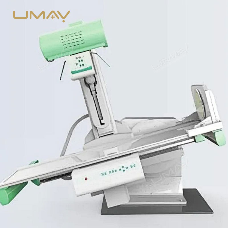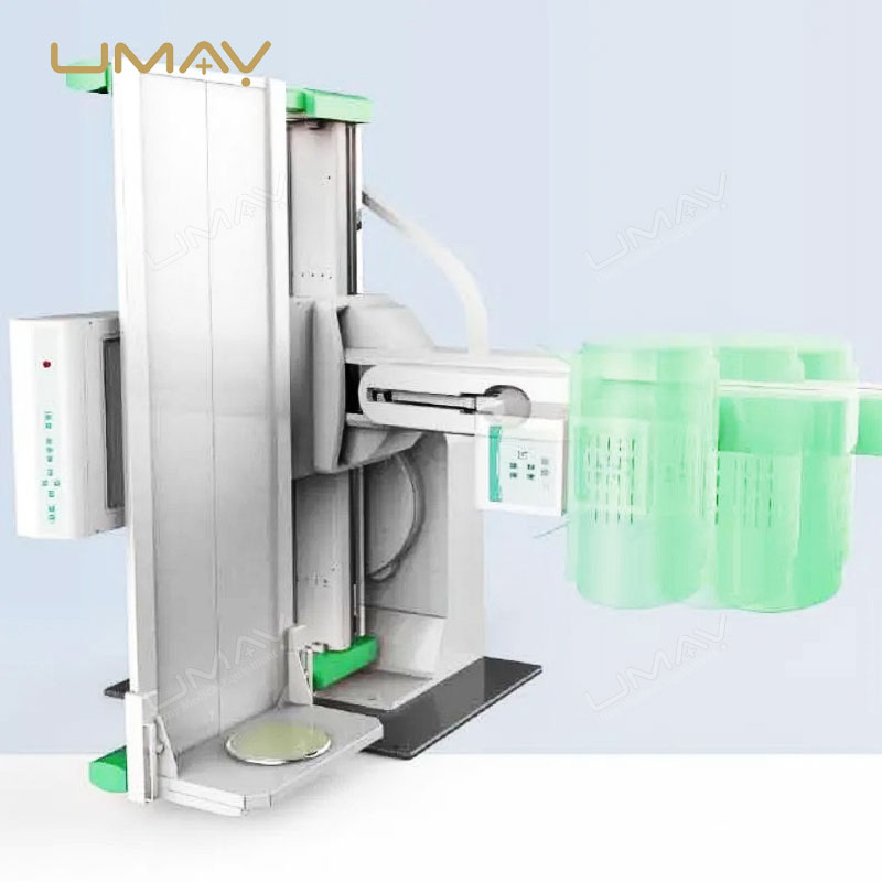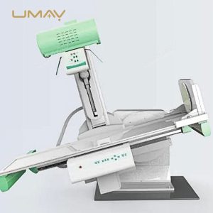

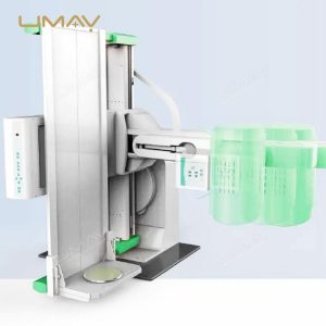


Advanced Digital Radiography and Fluoroscopy (DRF) System for High-Precision Imaging
SKU: UMY-XM-045
Brand: Umy
MOQ: 1
Origin: China
Quick Info.
- SKU NO.: UMY-XM-045
- Device Classification: Class Ⅱ
- Warranty: 1 Year
- Power Source: Electric
- Transport Package: Carton or Wooden Cases
- Origin: China
- Material: Aluminum and magnesium alloys
- After-Sale Service: Online Technical Support
- Production Capacity: 500 Sets/Year
The advanced digital radiography and fluoroscopy (DRF) system is designed to provide high-quality, real-time imaging for a wide range of diagnostic applications. This high-end system integrates both radiography and fluoroscopy capabilities, offering versatility and precision in medical imaging. Its digital technology ensures fast image acquisition and processing, improving workflow efficiency and diagnostic accuracy in hospitals and specialized imaging centers. Ideal for complex procedures, the DRF system enhances both patient care and clinical outcomes.
The Specific Parameters
| ITEMS | PARAMETERS |
|---|---|
| Power Supply | |
| Voltage | 380V ± 38V |
| Frequency | 50Hz ± 1Hz |
| Capacity | ≥65kVA |
| Internal Resistance | ≤0.17Ω |
| Generator | |
| Power | 80kW |
| Inverter Frequency | 440kHz |
| Radiography Tube Voltage | 40kV ~ 150kV step adjustment |
| Radiography Tube Current | 10mA ~ 1000mA step adjustment |
| Radiography Time | 1.0ms ~ 10,000ms step adjustment |
| Radiography mAs | 0.1mAs ~ 1000mAs |
| Fluoroscopy Tube Voltage | 40kV ~ 125kV step adjustment |
| Fluoroscopy Tube Current | 0.5mA ~ 10mA (continuous fluoroscopy), 5mA ~ 20mA (pulse fluoroscopy) |
| X-Ray Tube Assembly | |
| Target | Molybdenum-based lanthanum-tungsten composite |
| Target Angle | 12° |
| Nominal Tube Voltage | 150kV |
| Tube Focus (Big/Small) | 1.2mm / 0.6mm |
| Input Power | Big Focus: 100kW, Small Focus: 40kW |
| Anode Thermal Capacity | 420kJ (600kHU) |
| Anode Maximum Heat Dissipation | 1750W (2465kHU/min) |
| Component Heat Capacity | 1420kJ (2000kHU) |
| Anode Rotation Speed | 9700rpm |
| Collimator | |
| Field of View Light | Halogen lamp, AC24V/100W |
| Visible Light Illumination | Average illumination brightness > 100LUX |
| Light Field Exposure Time | 5 ~ 45s, 5s per step |
| Diagnostic Table | |
| Table Transverse Movement Distance | 300mm |
| SID | 1100mm ~ 1800mm |
| Table Panel Equivalent Filtration | ≤1.2mmAl |
| Point Device Movement Range | 1250mm |
| Table Height from Ground | 905mm |
| Table Size | 2100mm × 730mm |
| Table Rotation | +90° ~ -25° |
| X-Ray Tube Column Swing | +40° ~ 0° ~ -40° |
| Minimum Distance of Compressor from Table Surface | ≤150mm |
| Compressor Pressure | 80N ~ 130N |
| Table Bearing | 135kg |
| Fragmentation | Whole film, two-piece, four-slice |
| Fixed Grid | |
| Grid Density | 103L/INCH |
| Ratio | 0.41736111111111 |
| Convergence Distance | 130cm |
| Stationary Type | 15″ × 18″ |
| Intensifier | |
| Field of View Size | 230mm |
| Resolution | 52 Lp/cm |
| Conversion Factor | 26 |
| Signal-to-Noise Ratio | 45 dB |
| X-Ray Camera | |
| Cell Size | 5.86 × 5.86µm |
| Number of Valid Pixels | 1920 × 1200 |
| Flat Panel Detector | |
| Image Sensor | Amorphous silicon thin-film transistor |
| Pixel Size | 143µm |
| Effective Pixel Size | 3008(H) × 3072(V) |
| Effective Area (H × V) | 430(H) × 439(V) |
| Gray Scale | 14-bit |
| Spatial Resolution | 3.7 Lp/mm (CsI) |
| Energy Range | 40kV ~ 150kVP |
| Power Input | DC24V |
